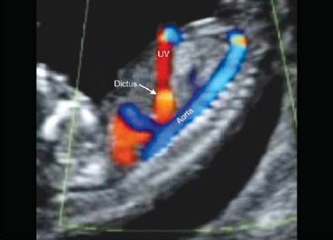
This scan is carried out from 11 weeks and 4 days to 13 weeks and 6 days. The best time (taking into account the small size of the fetus) is having the scan between 12 weeks and 3 days and 13 weeks 6 days. At this stage the fetal organs can also be examined and the gender determined in ± 70% of cases! A trans-abdominal scan is usually performed; however, it may be required to have a closer look vaginally.
The aim of the 11-14 Week scan (NT Scan)
- To date the pregnancy accurately (if this has not been done yet).
- To diagnose the type of twin (or other multiple) pregnancy. It is important to know whether each twin has its own placenta, or whether they share a common placenta. If they share a placenta, it is advisable to monitor the pregnancy more closely.
- To check whether the baby is growing and developing normally. Some major abnormalities can be visible at 12 weeks, but it is much better to have an ultrasound examination at 20 weeks to exclude structural abnormalities.
- To assess the risks of Down’s syndrome and other chromosomal abnormalities.
- Your individual risk for this pregnancy is calculated by taking into account your age, measurement of hormones in your blood and the ultrasound findings.
More about the 11-14 Week scan (NT Scan)
The size of the fetus is about 5-6.3 cm and weigh 14g.
Weeks 12-18 are a major, critical period in your baby’s brain development. Avoid any alcohol, tobacco or drugs because these could have long-term effects on your baby. This is also when your baby’s pituitary gland starts working and making its own hormones. Your baby’s placenta is now doing all the important work – filtering oxygen and nutrients to assist your baby’s growth. This week, your baby can suck its thumb. Its eyelids will fully cover the eyes, so they are well protected. Your baby’s intestines are starting to contract and relax, practicing for healthy digestion when they are born.
Structure which we measure/evaluate during screening for chromosomal abnormalities are:
- the fetal heart rate
- the nasal bone
- collection of fluid behind the fetal neck (nuchal translucency)
- the blood flow across the ductus venosus and
- tricuspid valve of the fetal heart (dependent on the fetal lie).
The Factors Assessed at the 1st Trimester Nt Scan are summarised as
Age of the mother, amount of fluid behind the neck of the fetus, presence or absence of the fetal nasal bone, fetal heart rate, blood flow through the tricuspid valve of the fetal heart, blood flow through the ductus venosus in the fetal liver and the level of 2 proteins in the mother’s blood (free B-hCG and PAPP-A). This blood test is known as the first trimester Downs syndrome biochemistry. The blood test is ideally done at 10 weeks; however, it can be done between 8 weeks and 0 days and 13 weeks and 6 days.

With the results of the blood test included in the calculation, the detection rate for Downs syndrome is approximately 95% and without the blood test results, 80%. Not all babies with Downs syndrome will show physical signs on ultrasound and we therefor feel it is important to have the blood test done. At the end of the test a risk number is calculated. A risk number of 1:2 to 1:300 is high risk, 1:300 – 1:2 500 is intermediate risk and a number above 1:2 500 is low risk for Downs syndrome. The higher the number the less likely it is statistically that the baby has Downs syndrome.
There are other tests available that have a better than 95% detection rate for Downs Syndrome, but they are optional and only necessary if the patient screen intermediate or high risk.
- NIPT (Non-invasive prenatal testing) is a DNA test on maternal blood (therefor carries no risk of miscarriage) to screen pregnancies for the most common fetal chromosome anomalies: trisomy 21 (Down syndrome), trisomy 18 (Edwards syndrome) and trisomy 13 (Patau syndrome). Also, the gender of the fetus is determined. The NIPT is highly sensitive and picks up more than 99% of cases of Down syndrome. But it is still a screening test rather than a diagnostic test. Unfortunately, this test is still expensive in South Africa, approximately R6 500 – R7 500.
- At the current time, karyotype testing in the prenatal setting is quite invasive, requiring amniocentesis or chorionic villus sampling and carries a small risk of miscarriage, approximately 1 in 300. Karyotyping can be done from blood, hair, or any other tissue. However, most karyotyping for medical diagnostic purposes is done on embryonic or fetal cells from unborn babies still in the uterus. The cells are usually collected by one of two methods: amniocentesis (amniotic fluid collected) performed after 16 weeks or chorionic villi sampling (piece of the placenta collected) performed between 12-14 weeks. The detection rate for Down’s syndrome is 99.99% and we therefor call it a diagnostic test.
| Method of screening | Proportion of all cases of T21 |
|---|---|
| Maternal age | 30%/50% |
| Age, NT, FHR, B-hCG, PAPPA-A | 90% |
| Second Trimester serum biochemistry | 50-70% |
| Combined plus additional ultrasound markers | 95% |
| Cell Free DNA in maternal blood | >99% |
* Courtesy of FMF
It is important that an accredited health care professional (might it be a Sonographer, Sonologist, Obstetrician or Fetal Medicine specialist) conduct the first trimester screening. To avoid disappointment, please confirm in advance on the Fetal Medicine Foundation (FMF) if the person is accredited.

More about the 11-14 Week scan (NT Scan)
The size of the fetus is about 5-6.3 cm and weigh 14g.
Weeks 12-18 are a major, critical period in your baby’s brain development. Avoid any alcohol, tobacco or drugs because these could have long-term effects on your baby. This is also when your baby’s pituitary gland starts working and making its own hormones. Your baby’s placenta is now doing all the important work – filtering oxygen and nutrients to assist your baby’s growth. This week, your baby can suck its thumb. Its eyelids will fully cover the eyes, so they are well protected. Your baby’s intestines are starting to contract and relax, practicing for healthy digestion when they are born.